Research Disclaimer: Testosterone esters and gonadotropins are sold exclusively for in-vitro and laboratory research use. Not for unsupervised use. This content does not constitute medical advice.
Exogenous testosterone administration suppresses the hypothalamic-pituitary-testicular (HPT) axis through negative feedback — one of the most important and well-studied consequences of supraphysiological androgen exposure. Understanding the mechanisms, timeline, and reversibility of this suppression is critical for research involving androgen replacement, male contraception, and gonadotropin biology.
Table of Contents#
- The HPTA: Normal Axis Function
- Negative Feedback Mechanisms
- Gonadotropin Suppression Timeline
- Testicular Atrophy Mechanism
- Spermatogenesis Suppression
- Dose-Dependence of Suppression
- HPTA Recovery After Cessation
- Research Models for HPTA Study
- Restoring HPTA: hCG, SERMs, and GnRH Analogues
- Research Protocols
- Key References
The HPTA: Normal Axis Function#
The hypothalamic-pituitary-testicular (HPTA) axis is the central endocrine control system for male reproductive function and testosterone production:
Hypothalamus:
- Kisspeptin neurons (AVPV and ARC nuclei) → regulate GnRH pulse generator
- GnRH (gonadotropin-releasing hormone) → secreted in pulses (approximately every 60–90 minutes in males)
- Pulsatile GnRH secretion is essential — constant GnRH levels paradoxically suppress gonadotropins (GnRH agonist paradox)
Anterior pituitary:
- GnRH → gonadotroph cells → LH (luteinizing hormone) and FSH (follicle-stimulating hormone) released in pulses mirroring GnRH
- LH: drives Leydig cell testosterone production
- FSH: drives Sertoli cell function and spermatogenesis support
Testes:
- LH → Leydig cells → testosterone biosynthesis (CYP11A1 → 3β-HSD → CYP17A1 → 17β-HSD)
- FSH → Sertoli cells → spermatogenesis support (androgen-binding protein, inhibin B)
Negative Feedback Mechanisms#
Exogenous testosterone suppresses the HPTA at two levels:
1. Hypothalamic suppression (kisspeptin/GnRH pulse frequency)#
- Testosterone → AR on kisspeptin neurons (ARC) → reduced kisspeptin firing → reduced GnRH pulse frequency
- DHT (from 5α-reduction of testosterone) → AR → additional suppressive signal in ARC
- E2 (from aromatization of testosterone) → ERα on AVPV and ARC kisspeptin neurons → potent suppression of GnRH pulse amplitude and frequency
E2 is the most potent suppressor at the hypothalamic level: studies using aromatase inhibitors show that blocking testosterone aromatization significantly reduces HPTA suppression, revealing that estrogen (not testosterone itself) is responsible for a major fraction of negative feedback.
2. Pituitary suppression (gonadotropin secretion)#
- Testosterone → AR on gonadotroph cells → direct suppression of LH and FSH transcription and secretion
- E2 → ERα on gonadotroph cells → potent suppression of LH and FSH pulse amplitude
- GnRH receptor downregulation → with reduced GnRH input, pituitary gonadotroph GnRH receptor expression decreases → further reduces responsiveness to hypothalamic GnRH
Summary of suppression hierarchy#
| Hormone | Primary suppressive signal for HPTA | Secondary |
|---|---|---|
| Testosterone | Hypothalamic (AR on kisspeptin neurons) + Pituitary (AR on gonadotrophs) | Both levels |
| DHT | Hypothalamic (AR on GnRH interneurons) | — |
| E2 (from aromatization) | Hypothalamic (ERα on kisspeptin/AVPV) + Pituitary (ERα on gonadotrophs) | Dominant at both levels |
Gonadotropin Suppression Timeline#
After initiation of exogenous testosterone (e.g., TE 200 mg/week)#
| Timepoint | LH | FSH | Serum T |
|---|---|---|---|
| Baseline | ~2–8 IU/L | ~1–10 IU/L | ~400–800 ng/dL (intact males) |
| Day 3–5 | Declining | Declining | Rising (TE peak) |
| Week 1 | ~0.5–2 IU/L | ~1–5 IU/L | ~1500–2500 ng/dL |
| Week 2 | ~0.1–0.5 IU/L | ~0.5–2 IU/L | ~1000–1500 ng/dL |
| Week 4 (steady-state) | <0.1–0.3 IU/L (near suppressed) | <1–2 IU/L | ~1000–1800 ng/dL |
| Month 3+ (chronic) | Near 0 (<0.1 IU/L) | Near 0 or very low | Maintained by exogenous T |
LH suppresses faster than FSH: LH is more acutely sensitive to testosterone/E2 feedback; FSH has a longer half-life (~3 days vs. ~1 day for LH) and responds more slowly and less completely to androgens alone (FSH is also regulated by inhibin B from Sertoli cells).
Testicular Atrophy Mechanism#
With prolonged HPTA suppression, the testes atrophy due to the absence of trophic gonadotropin stimulation:
Leydig cell changes#
- Without LH: Leydig cells lose their primary trophic signal
- Leydig cells atrophy: reduced cell volume, reduced StAR expression, reduced CYP enzyme expression
- Intratesticular testosterone (ITT) falls dramatically: ITT is normally 50–200× higher than serum testosterone (~20,000–50,000 ng/dL) — essential for spermatogenesis
- Even with exogenous testosterone maintaining serum levels ~1000 ng/dL, ITT can fall to near systemic levels (~1000 ng/dL) → insufficient for spermatogenesis
Sertoli cell changes#
- Without FSH + without high ITT: Sertoli cell function impaired
- Sertoli cells support germ cells throughout spermatogenesis — their dysfunction causes germ cell apoptosis
- Reduced Sertoli cell tight junctions (blood-testis barrier compromise) with long-term deprivation
Gross testicular atrophy#
- Testicular volume decreases proportionally to duration of suppression
- Typical: 20–30% volume decrease within 4–8 weeks of complete LH/FSH suppression
- Severe (12+ months): 50%+ volume reduction in some cases
Spermatogenesis Suppression#
Spermatogenesis requires:
- FSH → Sertoli cell activation
- High ITT (50–200× serum testosterone) → androgen-dependent germ cell progression
Exogenous testosterone suppresses BOTH:
- FSH drops to near zero → Sertoli cells lose FSH-driven support
- ITT falls drastically (despite high serum testosterone) → germ cells lose androgen support
Timeline to azoospermia (zero sperm count) with testosterone alone:
- Sperm count falls from ~20–200 million/mL to near-azoospermia in 12–16 weeks in most men
- Not 100% effective in all males (ethnic variation — lower efficacy in Caucasian males vs. Asian males, historically studied in WHO male contraception trials)
Dose-Dependence of Suppression#
Testosterone enanthate dose-response for LH suppression (human data)#
| TE dose (weekly) | LH at steady-state | FSH at steady-state |
|---|---|---|
| 25 mg/week | Partially suppressed (~50–70%) | Partially suppressed (~30–50%) |
| 100 mg/week | Severely suppressed (~80–90%) | Severely suppressed (~60–80%) |
| 200 mg/week | Near-complete (>95%) | Near-complete (>90%) |
| 600 mg/week | Complete (<0.1 IU/L) | Complete (<0.5 IU/L) |
Even physiological-range TRT (100 mg/week maintaining T ~500–800 ng/dL) produces significant LH/FSH suppression. Complete suppression requires supraphysiological levels.
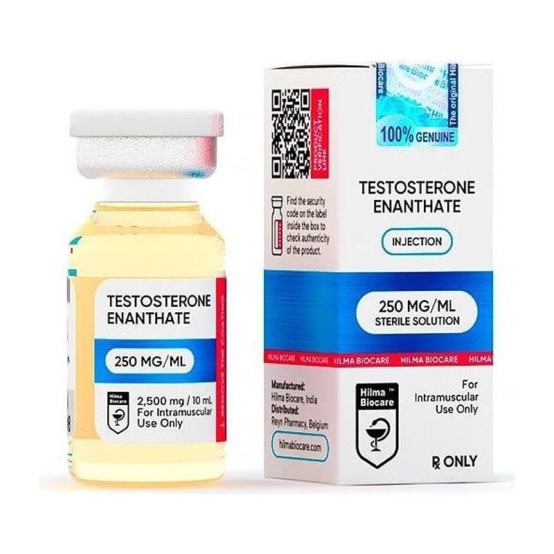
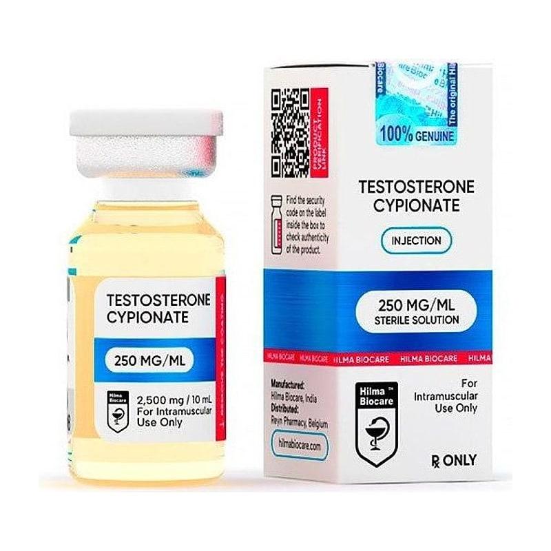
Testosterone Cypionate, €35 · 99.0% purityTestosterone Enanthate, €37 · 99.2% purity
HPTA Recovery After Cessation#
Recovery timeline after exogenous testosterone cessation:
| Timeframe (post-cessation) | LH/FSH | Endogenous T | Spermatogenesis |
|---|---|---|---|
| Weeks 1–4 | LH/FSH beginning to rise | Still low | Still suppressed |
| Month 1–2 | LH/FSH rising toward normal | T rising slowly | Oligospermia |
| Month 2–6 | LH/FSH normalizing | T approaching baseline | Recovering |
| Month 6–12 | Normal in most cases | Normal in most cases | Normal in most cases |
| >12 months (rare persistent) | Persistent hypogonadism | Below baseline | Incomplete |
Key variables determining recovery time:
- Duration of suppression (longer = slower recovery)
- Total dose (higher = slower recovery)
- Individual variation (pituitary gonadotroph reserve, testicular LH receptor density)
- Age (older males recover more slowly)
- Use of hCG or SERMs during recovery (accelerates recovery)
Research Models for HPTA Study#
Intact rodent model (high serum testosterone → HPTA suppression)#
| Parameter | Specification |
|---|---|
| Model | Adult intact male SD rats |
| Testosterone dose | 10 mg/kg TE SC every 3 days |
| Duration | 4–12 weeks |
| Endpoints | Serum LH, FSH, testosterone (trough), testicular weight, sperm count, ITT |
Castrated → LH/FSH measurement without testicular feedback#
Castration removes testicular testosterone production and testicular inhibin B:
- LH and FSH rise dramatically (50–200× above intact levels) within 7 days post-castration
- This allows studying hypothalamic and pituitary sensitivity to steroid hormone feedback in isolation
- Re-administering testosterone (various doses) to castrated animals → dose-response of gonadotropin suppression
Restoring HPTA: hCG, SERMs, and GnRH Analogues#
hCG (human chorionic gonadotropin)#
hCG is structurally homologous to LH (both bind LH receptor):
- hCG → Leydig cells (bypasses HPT suppression) → directly stimulates intratesticular testosterone production
- hCG prevents or reverses Leydig cell atrophy during or after suppression
- Co-administration with testosterone prevents ITT decline even during full HPTA suppression
Research protocol: hCG 500 IU SC every 3 days co-administered with testosterone maintains ITT at supraphysiological levels → preserves testicular volume and Leydig cell function.
SERMs (tamoxifen, clomiphene)#
SERMs act as ER antagonists at hypothalamus and pituitary:
- Block E2's negative feedback at hypothalamic ERα (kisspeptin neurons) and pituitary ERα (gonadotrophs)
- Result: increased GnRH pulse frequency → increased LH/FSH → increased endogenous testosterone
- Used for HPTA restart research post-suppression
GnRH pulse therapy#
Pulsatile GnRH administration (mimicking normal pulse frequency via mini-pump) can restore LH/FSH secretion even after prolonged HPTA suppression — demonstrating that the pituitary gonadotrophs are not intrinsically damaged, only functionally suppressed.
Research Protocols#
HPTA suppression and recovery study#
| Phase | Protocol |
|---|---|
| Baseline (Week -2 to 0) | Serum LH, FSH, T, sperm count. Testicular volume |
| Suppression phase (Weeks 1–16) | TE 200 mg/week IM. Blood draws at weeks 1, 2, 4, 8, 12, 16 |
| Cessation (Week 16 onward) | Stop TE. Blood draws at weeks 17, 18, 20, 24, 28, 32 |
| Endpoints | LH, FSH, total T, free T, E2, SHBG, inhibin B, sperm count, testicular volume |
Dose-response gonadotropin suppression study (rodents)#
| Parameter | Specification |
|---|---|
| Model | Adult intact male SD rats |
| TE doses | 0, 1, 3, 10, 30 mg/kg SC |
| Frequency | Every 3 days |
| Duration | 6 weeks |
| Endpoints | Serum LH (ELISA), FSH (ELISA), testosterone, testicular weight, ITT by LC-MS/MS |
Key References#
-
Contraception (2000). WHO Task Force on Methods for the Regulation of Male Fertility. "Contraceptive efficacy of testosterone-induced azoospermia." Lancet, 336:955–959.
-
Hayes FJ, et al. (2001). "Testosterone-induced increase in muscle size in healthy young men is associated with muscle fiber hypertrophy." American Journal of Physiology-Endocrinology and Metabolism, 281:E1169–E1178.
-
Swerdloff RS, et al. (2000). "Long-term pharmacokinetics of transdermal testosterone gel in hypogonadal men." Journal of Clinical Endocrinology & Metabolism, 85(12):4500–4510.
-
Veldhuis JD, et al. (2001). "Testosterone supplementation in older men restrains insulin-like growth factor-I (IGF-I)-induced oligoclonal expansion of T-lymphocytes." Journal of Clinical Endocrinology & Metabolism, 86:5536–5545.
Research Use Only: All compounds listed are sold exclusively for in-vitro research and laboratory use. Not for unsupervised use. JuicedPharma does not provide medical advice.
Published by JuicedPharma Research Team · September 2026 · Category: Research Protocols & Science