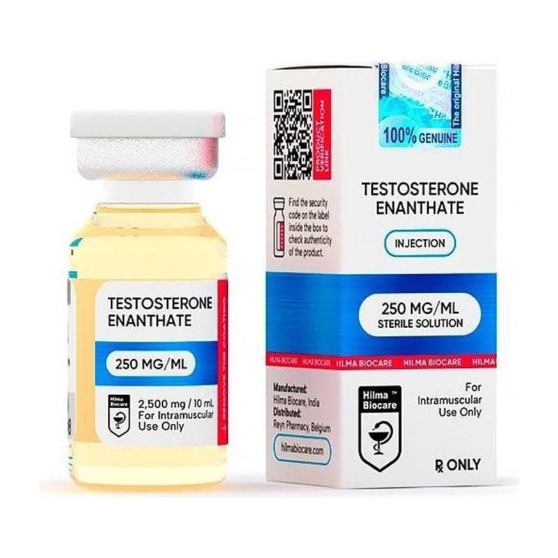
Research Disclaimer: Testosterone enanthate is sold exclusively for in-vitro and laboratory research use. Not for unsupervised use. This content does not constitute medical advice.
Testosterone enanthate (TE) is the C17β-enanthate ester of testosterone — the most widely used long-acting injectable testosterone in both clinical medicine (testosterone replacement therapy, hypogonadism treatment) and research settings. Its well-characterized pharmacokinetics, decades of published data, and predictable IM absorption profile make it the reference standard for studying androgen receptor biology, anabolic signaling, and sex hormone physiology.
Table of Contents#
- Testosterone: The Primary Male Androgen
- Enanthate Ester: Pharmacokinetic Rationale
- Pharmacokinetics After IM Injection
- Androgen Receptor Signaling
- Anabolic Mechanisms: Muscle Protein Synthesis
- Satellite Cell Activation and IGF-1 Crosstalk
- Androgenic Effects and 5α-Reduction
- Aromatization and Estrogenic Effects
- HPTA Suppression
- Research Protocols
- Key References
Testosterone: The Primary Male Androgen#
Testosterone is a C19 steroid hormone produced primarily by testicular Leydig cells (in males) and in smaller amounts by the adrenal cortex and ovaries (in females).
Biosynthesis pathway: Cholesterol → pregnenolone → DHEA → androstenedione → testosterone (Key enzymes: CYP11A1, 3β-HSD, CYP17A1, 17β-HSD)
Normal circulating levels:
- Adult males: 300–1000 ng/dL (10.4–34.7 nmol/L) total testosterone
- Free testosterone (~2–3%): 50–200 pg/mL (the biologically active fraction not bound to SHBG or albumin)
Testosterone's mechanisms of action:
- Direct AR activation: testosterone binds androgen receptor (AR, nuclear receptor) → AR-mediated gene expression
- 5α-reduction to DHT: in certain tissues (prostate, skin, scalp), 5α-reductase converts testosterone to the more potent dihydrotestosterone
- Aromatization to E2: aromatase (CYP19A1) in adipose, liver, brain, gonads converts testosterone to estradiol (E2) — important for bone health, libido, cardiovascular effects
Enanthate Ester: Pharmacokinetic Rationale#
Why esterify testosterone?
Unmodified (native) testosterone:
- Very short half-life after IM injection: ~0.5–1 hour (rapidly absorbed and cleared)
- Impractical for chronic research or therapeutic use (requires frequent dosing)
Esterification at the 17β-hydroxyl group slows absorption:
- Esterified testosterone is oil-soluble — injected in oil vehicle (sesame, MCT) → depot forms at injection site
- Ester must be cleaved by esterases (non-specific esterases ubiquitous in plasma and tissue) before testosterone is bioactive
- Ester hydrolysis rate determines t½ — longer ester chain = slower hydrolysis = longer t½
Enanthate ester: 7-carbon chain → intermediate length:
- t½ after IM injection: ~4.5 days
- Dosing interval in research/clinical use: typically once weekly (maintains stable levels)
Pharmacokinetics After IM Injection#
In humans (clinical data)#
Testosterone enanthate IM injection:
- Absorption: from oil depot over ~10–14 days
- Cmax: ~1500–2500 ng/dL at 24–72h post-injection (standard 200 mg dose)
- t½: ~4.5 days (effective t½ depends on absorption from depot)
- Steady-state (weekly dosing): reached after 3–4 weeks
- Return to baseline: ~14–21 days after last injection
In rodents#
Rodent pharmacokinetics differ substantially:
- Higher metabolic rate → faster testosterone clearance
- t½ shorter in rodents (~1–2 days)
- For weekly injections: testosterone levels fluctuate more than in humans
- Common research approach: 3× weekly injections in rodents for smoother curves; or daily SC
Castrated rodent model (orchiectomy + TE replacement)#
The gold standard for studying androgen receptor biology:
- Castrate animals (bilateral orchiectomy) → removes endogenous testosterone
- Administer TE at defined doses → replace androgen at controlled levels
- Allows dose-response studies without confounding from endogenous production
- Commonly used: 0.5–5 mg/kg SC every 3 days in rats; 10 mg/kg SC weekly in mice
Androgen Receptor Signaling#
AR structure and activation#
The androgen receptor (AR, NR3C4) is a member of the nuclear receptor superfamily:
- Ligand-binding domain (LBD): C-terminal; binds testosterone, DHT
- DNA-binding domain (DBD): zinc fingers; binds androgen response elements (ARE) in gene promoters
- N-terminal domain (NTD): transactivation function AF-1; N-C (NTD-LBD) interaction required for full activation
Canonical genomic AR signaling:
- Testosterone or DHT enters cell → binds AR in cytoplasm (complexed with Hsp90 chaperone)
- Testosterone/AR → conformational change → Hsp90 dissociation → AR homodimerization → nuclear translocation
- AR homodimer → binds ARE (TGTTCT repeat) in target gene promoters
- AR → recruits coactivators (SRC-1, CBP/p300) → histone acetylation → gene transcription
Key AR target genes in muscle:
- Insulin-like growth factor binding protein-3 (IGFBP-3)
- Follistatin (inhibits myostatin/activin signaling)
- Androgen-regulated genes in protein synthesis (S6K1, eIF4E targets)
- IGF-1 (in liver and muscle — AR-dependent IGF-1 expression in skeletal muscle)
Anabolic Mechanisms: Muscle Protein Synthesis#
Testosterone's anabolic effect in skeletal muscle is well established and mediated through multiple mechanisms:
1. Direct AR-mediated transcription in muscle#
Muscle fibers express AR on myonuclei. Testosterone → AR → ARE-driven gene expression:
- Increased muscle-specific IGF-1 (mIGF-1/IGF-1Ea) mRNA
- Increased follistatin → reduced myostatin inhibition → enhanced myogenesis
- Increased mTOR pathway genes
2. PI3K/Akt/mTOR pathway#
Beyond nuclear AR effects, testosterone also activates membrane-associated signaling:
- Testosterone → membrane AR (non-genomic) → PI3K activation → Akt → mTOR → p70S6K → protein synthesis
- This non-genomic pathway is faster than nuclear AR signaling (minutes vs. hours)
3. Anti-proteolytic effects#
Testosterone reduces ubiquitin-proteasome pathway activity in muscle:
- Reduces MAFbx (muscle atrophy F-box, atrogin-1) and MuRF1 expression
- These E3 ubiquitin ligases are responsible for muscle protein degradation during atrophy
- AR-mediated reduction in their expression → reduced protein breakdown
Net result: greater net protein balance (synthesis > breakdown) → muscle hypertrophy.
Satellite Cell Activation and IGF-1 Crosstalk#
Testosterone promotes muscle stem cell (satellite cell) activation:
- AR is expressed on satellite cells
- Testosterone → AR → follistatin expression in satellite cells → reduced myostatin (myostatin is the most potent negative regulator of satellite cell activation)
- Reduced myostatin → satellite cells enter cell cycle → proliferate → fuse with existing fibers → myonuclear addition → increased myonuclear domain capacity → enhanced protein synthesis per fiber
IGF-1 crosstalk: Testosterone → hepatic and muscle IGF-1 production → IGF-1 → satellite cell and myofiber IGF-1R → PI3K/Akt → mTOR. This creates an autocrine/paracrine amplification loop: testosterone → IGF-1 → more anabolism → more AR expression (IGF-1 increases AR expression in muscle) → more testosterone sensitivity.
Androgenic Effects and 5α-Reduction#
In androgen-sensitive tissues (prostate, skin, hair follicles):
- 5α-reductase type 1 (skin) and type 2 (prostate, scalp) converts testosterone → DHT
- DHT has 3–5× higher AR binding affinity than testosterone (slower dissociation rate from AR)
- DHT cannot be aromatized to estrogen → purely androgenic
For research comparing anabolic vs. androgenic effects:
- 5α-reductase inhibitors (finasteride — blocks type 2; dutasteride — blocks both) allow studying what testosterone effects are DHT-dependent
- Tissue: ventral prostate weight → classic androgenic endpoint in castrated rat
- Levator ani muscle weight → anabolic endpoint (high AR, low 5α-reductase) in castrated rat
Aromatization and Estrogenic Effects#
Aromatase (CYP19A1)#
Aromatase converts testosterone to estradiol (E2) via aromatization of ring A:
- Primary aromatase sites: adipose tissue, liver, brain, bone, testis
- Aromatase activity increases with increasing adiposity → more E2 in obese males
E2 derived from testosterone — research implications#
In males:
- E2 derived from testosterone is required for bone mineral density maintenance (AR-independent)
- E2 for libido (brain aromatization → estrogen receptor activation in hypothalamus)
- E2 contributes to negative feedback on HPTA (both AR-mediated and ER-mediated)
For research: when studying pure AR effects, aromatase inhibitors (anastrozole, letrozole) are used to prevent testosterone-to-estradiol conversion. This allows dissecting AR-mediated vs. ER-mediated testosterone effects.
HPTA Suppression#
Hypothalamic-pituitary-testicular axis (HPTA): Hypothalamus → GnRH (pulsatile) → pituitary → LH + FSH → testes → testosterone + spermatogenesis
Exogenous testosterone (as TE) → elevated circulating testosterone → negative feedback:
- At hypothalamus: testosterone + DHT + E2 → reduced GnRH pulse frequency and amplitude
- At pituitary: testosterone + E2 → direct suppression of LH and FSH secretion (gonadotroph cells)
- Net: LH and FSH approach zero → testicular Leydig cells without LH stimulation → endogenous testosterone production falls to near zero → testicular atrophy
In research models studying testosterone effects: castrated models are preferred because endogenous production is eliminated, allowing clean dose-response characterization. In intact animals, TE administration partially suppresses but does not eliminate endogenous production → mixed hormone levels.
Research Protocols#
Castrated rat anabolic/androgenic assay (Hershberger assay)#
| Parameter | Specification |
|---|---|
| Model | Adult male Sprague-Dawley rats, bilateral orchiectomy |
| TE dose range | 0.1–5 mg/kg SC |
| Vehicle | Oil (MCT or sesame oil) |
| Frequency | Every 3 days |
| Duration | 2–3 weeks |
| Endpoints | Levator ani weight (anabolic), ventral prostate weight (androgenic), seminal vesicle weight, serum LH, FSH, SHBG |
Hypogonadism replacement model#
| Parameter | Specification |
|---|---|
| Model | Castrated adult male C57BL/6J mice |
| Dose groups | 0.1 mg/kg (low), 0.5 mg/kg (mid), 2 mg/kg (high), sham surgery control |
| Route | SC injection |
| Frequency | Every 3 days |
| Duration | 4–8 weeks |
| Primary endpoints | Muscle mass (quadriceps, gastrocnemius), body composition, serum testosterone, insulin sensitivity |
Key References#
-
Bhasin S, et al. (2001). "Testosterone dose-response relationships in healthy young men." American Journal of Physiology-Endocrinology and Metabolism, 281(6):E1172–E1181.
-
Mooradian AD, et al. (1987). "Biological actions of androgens." Endocrine Reviews, 8(1):1–28.
-
Basaria S. (2010). "Androgen abuse in athletes: detection and consequences." Journal of Clinical Endocrinology & Metabolism, 95(4):1533–1543.
-
Sinha-Hikim I, et al. (2002). "Testosterone-induced muscle hypertrophy is associated with an increase in satellite cell number in healthy, young men." American Journal of Physiology-Endocrinology and Metabolism, 285(1):E197–E205.
Research Use Only: Testosterone enanthate is sold exclusively for in-vitro research and laboratory use. Not for unsupervised use. JuicedPharma does not provide medical advice.
Published by JuicedPharma Research Team · September 2026 · Category: Research Protocols & Science