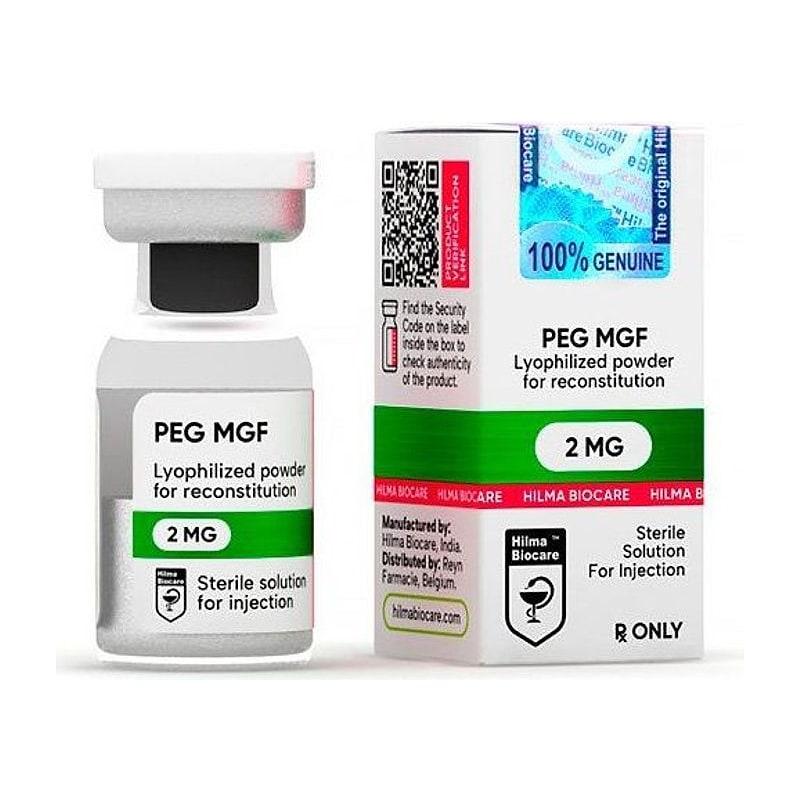
Research Disclaimer: PEG-MGF is sold exclusively for in-vitro and laboratory research use. Not approved for human therapeutic use. This content does not constitute medical advice.
Mechano Growth Factor (MGF) is an alternatively spliced variant of the IGF-1 gene that is specifically upregulated in response to mechanical stress in muscle — the body's local repair signal triggered by exercise or injury. PEG-MGF is the pegylated (polyethylene glycol-modified) form, engineered to extend the extremely short plasma half-life of native MGF from minutes to days while preserving its satellite cell-activating activity.
Understanding MGF's role in the satellite cell cycle is foundational for muscle repair research — it occupies a distinct niche from systemic GH/IGF-1 signaling that is specifically local and mechanically triggered.
Table of Contents#
- The IGF-1 Gene and Its Splice Variants
- What Makes MGF Unique: The E Domain
- Satellite Cells: The Muscle Stem Cell Population
- MGF's Role in the Satellite Cell Cycle
- PEGylation: Why MGF Needs It
- PEG-MGF vs. Native MGF in Research
- Comparison to Systemic IGF-1 and GH
- Research Data: Muscle Regeneration
- Protocol Design
- Frequently Asked Questions
- Key References
The IGF-1 Gene and Its Splice Variants#
The IGF-1 gene produces multiple protein isoforms through differential splicing of its six exons. The three main splice variants in muscle are:
-
IGF-1Ea (systemic IGF-1): the liver-produced, GH-responsive form that circulates to all tissues. Mediates most systemic anabolic effects of GH.
-
IGF-1Eb: expressed in liver and partially in muscle; function incompletely characterized.
-
IGF-1Ec (Mechano Growth Factor, MGF): expressed specifically in muscle in response to mechanical loading. Contains a unique E-peptide (the Ec extension peptide) at the C-terminus after splicing.
All three variants produce the same mature IGF-1 peptide domain after proteolytic processing, but the E peptide in each isoform is different and confers distinct receptor-binding and biological properties.
Why MGF is specifically a "mechanical" growth factor#
In unstressed muscle, IGF-1Ec expression is low. Within hours of mechanical stress (exercise-induced micro-damage, injury, or mechanical stretch in vitro):
- Local muscle IGF-1Ec mRNA is strongly upregulated
- The Ec E-peptide is released locally
- This local signal is distinct from systemic GH-driven IGF-1 (IGF-1Ea) production in the liver
This makes IGF-1Ec/MGF a local, mechanically-triggered repair signal — separate from the systemic GH/IGF-1 axis.
What Makes MGF Unique: The E Domain#
The defining feature of MGF is its Ec E-peptide — the 24-amino-acid C-terminal extension unique to the IGF-1Ec splice variant.
Structure of the Ec E-peptide#
The Ec E-peptide sequence (the fragment corresponding to the MGF research peptide) is: YQPPSTNKNTKSQRRKGSTFEEHK (human)
This peptide is released after proteolytic processing of the precursor MGF protein. The processed E-peptide has independent biological activity — distinct from the mature IGF-1 domain.
Why the E-peptide matters#
The IGF-1 domain in MGF signals through the canonical IGF-1R (same as systemic IGF-1). But the MGF E-peptide has additional distinct receptor interactions:
- Does not bind IGFBP-3 (the major circulating IGF-1 carrier protein) — makes it freely available locally
- Has independent mitogenic and satellite cell-activating properties not fully mediated by IGF-1R
- May interact with specific cell surface receptors on satellite cells that are distinct from IGF-1R
The research peptide sold as "MGF" or "PEG-MGF" is typically the 24-amino-acid Ec E-peptide, not the full MGF precursor protein. Its activity is mediated specifically by the Ec E-peptide, not by the IGF-1 domain.
Satellite Cells: The Muscle Stem Cell Population#
To understand MGF's function, you need to understand satellite cells.
What are satellite cells?#
Satellite cells are the resident stem cells of adult skeletal muscle — a quiescent population of myogenic progenitor cells located between the sarcolemma and the basal lamina of muscle fibers. They are called "satellite" cells because of this anatomical position.
Their role in muscle repair#
Adult skeletal muscle has limited self-renewal capacity for post-mitotic myofibers. When muscle is damaged (by exercise, injury, or disease):
- Quiescent satellite cells are activated (MGF is one of the key activation signals)
- Activated satellite cells proliferate (Myf5+, MyoD+ — myogenic determination genes expressed)
- A subset of activated satellite cells undergo apoptosis or re-enter quiescence (self-renewal to maintain the satellite cell pool)
- Committed myoblasts differentiate (myogenin expression) and fuse with damaged fibers or with each other to form new myotubes/myofibers
This satellite cell activation → proliferation → differentiation cascade is the fundamental mechanism of muscle repair. MGF acts at the activation step.
MGF's Role in the Satellite Cell Cycle#
Activation of quiescent satellite cells#
Published research (Yang and Goldspink, 2002 and subsequent):
- MGF E-peptide stimulates satellite cell activation from quiescence into the cell cycle
- This is the distinct contribution of MGF vs. systemic IGF-1: systemic IGF-1 drives proliferation and differentiation of already-activated cells, but MGF specifically triggers the initial quiescence → activation transition
- In MGF-knockout conditions, fewer satellite cells are activated after muscle damage → impaired repair
Paracrine action#
MGF's extremely short plasma half-life (2–3 minutes) means it acts locally — produced by the damaged fiber, it signals to adjacent satellite cells. This paracrine, local action is the intended physiological function.
Interaction with systemic IGF-1#
MGF and systemic IGF-1 have complementary, sequential roles:
- MGF (local, early): activates satellite cells from quiescence (first 24–72h after injury)
- Systemic IGF-1 (GH-driven): drives proliferation and differentiation of activated satellite cells (ongoing through repair)
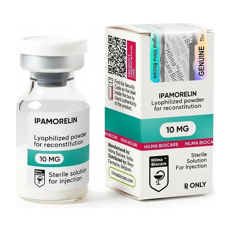
Optimal muscle repair uses both: local MGF activation triggers the satellite cell pool; systemic GH/IGF-1 provides the sustained anabolic drive for proliferation and hypertrophy. This is the rationale for combining PEG-MGF with GH secretagogues (Ipamorelin + CJC-1295) in recovery research protocols.
PEGylation: Why MGF Needs It#
The half-life problem#
Native MGF (the Ec E-peptide) has a plasma half-life of 2–5 minutes in serum due to rapid proteolytic degradation by plasma and tissue peptidases.
This ultra-short half-life is physiologically appropriate for a local paracrine signal (the body wants local action, not systemic spread). But it is a major problem for systemic research administration — a SC injection of native MGF would be degraded long before reaching distant muscle tissue.
PEGylation solution#
PEGylation is the attachment of polyethylene glycol (PEG) polymer chains to the peptide. The effects:
- Steric shielding: PEG chains form a hydrophilic "cloud" around the peptide → proteases cannot access cleavage sites → degradation slows dramatically
- Increased hydrodynamic radius: the PEG-peptide complex behaves as if it were a much larger molecule → renal filtration is reduced (smaller peptides are rapidly cleared by the kidneys)
- Reduced immunogenicity: PEG shielding reduces immune recognition of the foreign peptide
Result: PEG-MGF plasma half-life is approximately 72 hours (3 days), compared to 2–5 minutes for native MGF — a ~1000-fold extension.
PEGylation tradeoffs#
The PEG chain is biologically inert but introduces some considerations:
- Slightly reduced receptor binding affinity at the E-peptide receptor (PEG steric hindrance)
- Slower onset of action (absorption from SC depot is slower for larger PEGylated molecules)
- Research comparisons to native MGF require accounting for PK differences
PEG-MGF vs. Native MGF in Research#
| Parameter | Native MGF (Ec E-peptide) | PEG-MGF |
|---|---|---|
| Plasma half-life | 2–5 minutes | ~72 hours |
| Dosing frequency | Multiple/day (practical limit) | 1–2×/week |
| SC bioavailability | Very limited | Good |
| Receptor binding | Full affinity | Slightly reduced |
| Satellite cell activation | Yes (in vitro confirmed) | Yes (in vivo confirmed) |
| Systemic distribution | Very limited (local) | Widespread |
| Research application | In vitro, local injection protocols | In vivo, systemic administration |
For in vivo systemic research (studying effects on muscle throughout the body), PEG-MGF is the practical choice. For in vitro studies or local injection experiments, native MGF can be used.
Comparison to Systemic IGF-1 and GH#
| Parameter | PEG-MGF (Ec E-peptide) | Systemic IGF-1 | rhGH |
|---|---|---|---|
| Primary action | Satellite cell activation | Satellite cell proliferation/differentiation | IGF-1 production + direct effects |
| Timing in repair | Early (0–72h post-injury) | Ongoing (proliferation phase) | Ongoing |
| IGF-1R binding | Yes (IGF-1 domain) | Yes | Indirect (via IGF-1 production) |
| E-peptide receptor | Yes (unique MGF action) | No | No |
| Systemic IGF-1 elevation | Minimal | Yes | Yes |
| Lean mass (acute injury) | Strong (satellite cells) | Moderate | Indirect |
| Fat loss | None significant | Some | Yes (direct) |
PEG-MGF occupies a unique mechanistic position — it targets the muscle repair initiation step (satellite cell activation) that neither systemic IGF-1 nor GH directly drives. This makes it complementary to, rather than redundant with, GH secretagogues in recovery research.
Research Data: Muscle Regeneration#
Acute muscle injury models (rodent)#
Cardiotoxin injury, freeze injury, or bupivacaine injection (standard myotoxic injury models):
- PEG-MGF (1–2 mg/kg SC) administered within 24h of injury
- Endpoints: satellite cell number by immunostaining (Pax7+, MyoD+), myofiber cross-sectional area at 7/14/21 days, force production recovery
- Published results: faster satellite cell activation, more myotubes at day 7, greater fiber cross-sectional area at day 21 vs. vehicle
Aging models#
Muscle regenerative capacity declines with age due to reduced satellite cell activation in response to injury. In aged rodent models:
- PEG-MGF normalizes satellite cell activation rates closer to young animal levels
- Blunted, but real, improvement in muscle repair metrics
Combination with Ipamorelin + CJC-1295#
The rationale: PEG-MGF activates satellite cells → Ipamorelin + CJC-1295 elevates systemic GH/IGF-1 → the activated satellite cells receive a sustained anabolic drive for proliferation and hypertrophy. The combination should theoretically produce better repair than either component alone — though head-to-head combination data is limited.
Protocol Design#
Standard acute injury + repair protocol#
| Parameter | Specification |
|---|---|
| Model | Cardiotoxin-injected rat tibialis anterior |
| PEG-MGF dose | 1 mg/kg SC, once, within 12h of injury |
| Ipamorelin (co-treatment) | 100 µg/kg SC 2× daily × 14 days |
| Primary endpoints | Pax7+ satellite cells (day 3), CSA at days 7, 14, 21 |
| Secondary endpoints | Force production, fiber type distribution |
Overloaded muscle (hypertrophy model)#
Synergist ablation (removing the synergist muscle to force compensation hypertrophy):
- PEG-MGF 0.5–1 mg/kg SC twice weekly × 4 weeks
- Endpoint: muscle mass, satellite cell incorporation, CSA of hypertrophied fibers
Frequently Asked Questions#
Is PEG-MGF the same as IGF-1? No. PEG-MGF contains the Ec E-peptide sequence unique to the IGF-1Ec splice variant. While the full MGF precursor contains the IGF-1 domain, the E-peptide (the research peptide) has independent activity. It activates satellite cells via mechanisms distinct from (though partly overlapping with) IGF-1R signaling.
Why not just use IGF-1 instead of PEG-MGF? IGF-1 promotes satellite cell proliferation and differentiation (later phases), but MGF (via its E-peptide) specifically activates quiescent satellite cells (the initiation phase). They have complementary and partially non-overlapping roles. For muscle repair initiation, MGF is the more specific tool.
How often should PEG-MGF be dosed? Given its ~72h half-life, twice weekly (Monday/Thursday) SC injections are the standard dosing interval for chronic research protocols.
Can PEG-MGF be combined with BPC-157 and TB-500? Yes — the mechanisms are non-overlapping. BPC-157 (FAK-paxillin tendon/connective tissue) and TB-500 (G-actin sequestration, migration) act at different steps and tissue types than PEG-MGF (satellite cell activation in skeletal muscle). A comprehensive recovery stack covering satellite cells + actin dynamics + connective tissue + GH axis is a rational multi-modal approach.
Key References#
-
Yang SY, Goldspink G. (2002). "Different roles of the IGF-I Ec peptide (MGF) and mature IGF-I in myoblast proliferation and differentiation." FEBS Letters, 522(1-3):156–160.
-
Goldspink G. (2005). "Mechanical signals, IGF-I gene splicing, and muscle adaptation." Physiology, 20:232–238.
-
Carpenter V, et al. (2008). "Mechano-growth factor reduces loss of cardiac function in acute myocardial infarction." Heart, Lung and Circulation 17(1):33–39.
-
Dluzniewska J, et al. (2005). "A strong neuroprotective effect of the autonomous C-terminal peptide of IGF-1 Ec (MGF) in brain ischemia." FASEB Journal, 19(13):1896–1898.
-
Hill M, Goldspink G. (2003). "Expression and splicing of the insulin-like growth factor gene in rodent muscle is associated with muscle satellite (stem) cell activation following local tissue damage." Journal of Physiology, 549(2):409–418.
Research Use Only: PEG-MGF and all products referenced are sold exclusively for in-vitro research and laboratory use. Not for human consumption. JuicedPharma does not provide medical advice.
Published by JuicedPharma Research Team · September 2026 · Category: Growth Hormone