Research Disclaimer: All compounds described are sold exclusively for in-vitro and laboratory research use. Not for unsupervised use. This content does not constitute medical advice.
C17α-alkylated oral anabolic steroids are a class of compounds modified at the C17 position to resist hepatic first-pass metabolism. This modification — primarily a C17α-methyl group — enables oral bioavailability by blocking 17β-hydroxysteroid dehydrogenase activity, but at the cost of significantly increased hepatotoxicity. Understanding the mechanisms, biomarkers, histopathology, and hepatoprotective strategies for C17α-alkylation drug-induced liver injury (DILI) is essential for research protocols involving these compounds.
Table of Contents#
- The C17α-Alkylation Chemistry
- Hepatotoxicity Mechanisms
- Biomarkers: Liver Enzyme Panels
- Histopathology Spectrum
- Compound-Specific Hepatotoxicity Profiles
- Hepatoprotective Strategies in Research
- Research Protocol Design
- Key References
The C17α-Alkylation Chemistry#
Why C17α-methylation is needed for oral bioavailability:
- Testosterone and most steroidal androgens: 17β-OH group → rapidly oxidized by 17β-HSD in intestinal wall and liver → inactive 17-keto compound
- First-pass extraction: >90% of testosterone removed on first hepatic pass
- C17α-methyl (or ethyl) → sterically blocks 17β-HSD from oxidizing 17β-OH → compound survives first-pass → oral bioavailability increases to 50–80%
The trade-off: The same C17α-methyl group that blocks metabolic inactivation also:
- Dramatically increases liver exposure concentration (high portal vein concentration × oral bioavailability)
- Inhibits bile salt export pump (BSEP) → cholestasis
- Generates reactive metabolites via CYP450 → oxidative stress
Key C17α-alkylated research compounds:
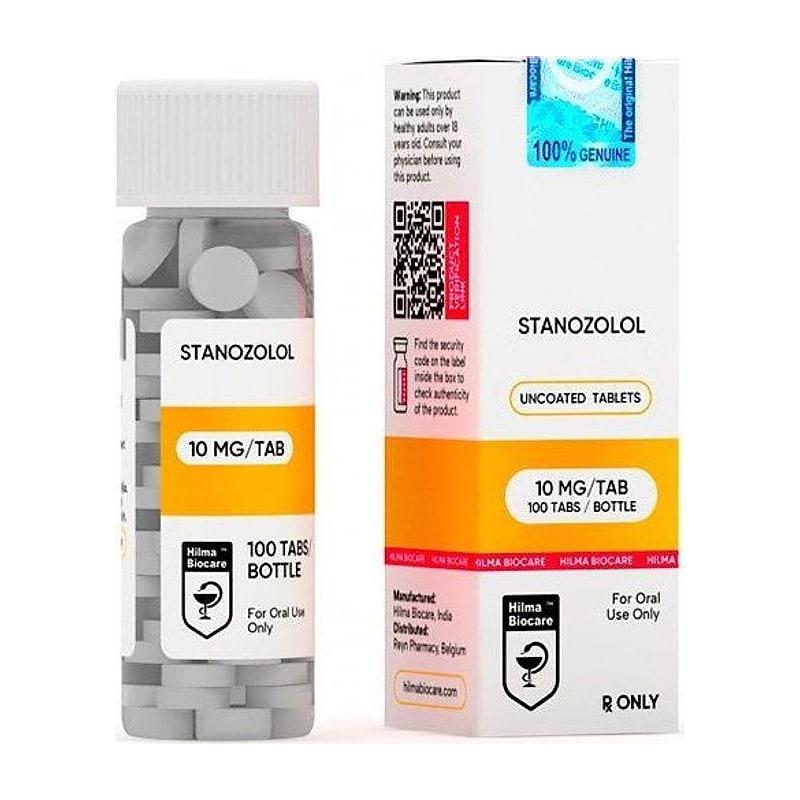
- Stanozolol (C17α-methyl)
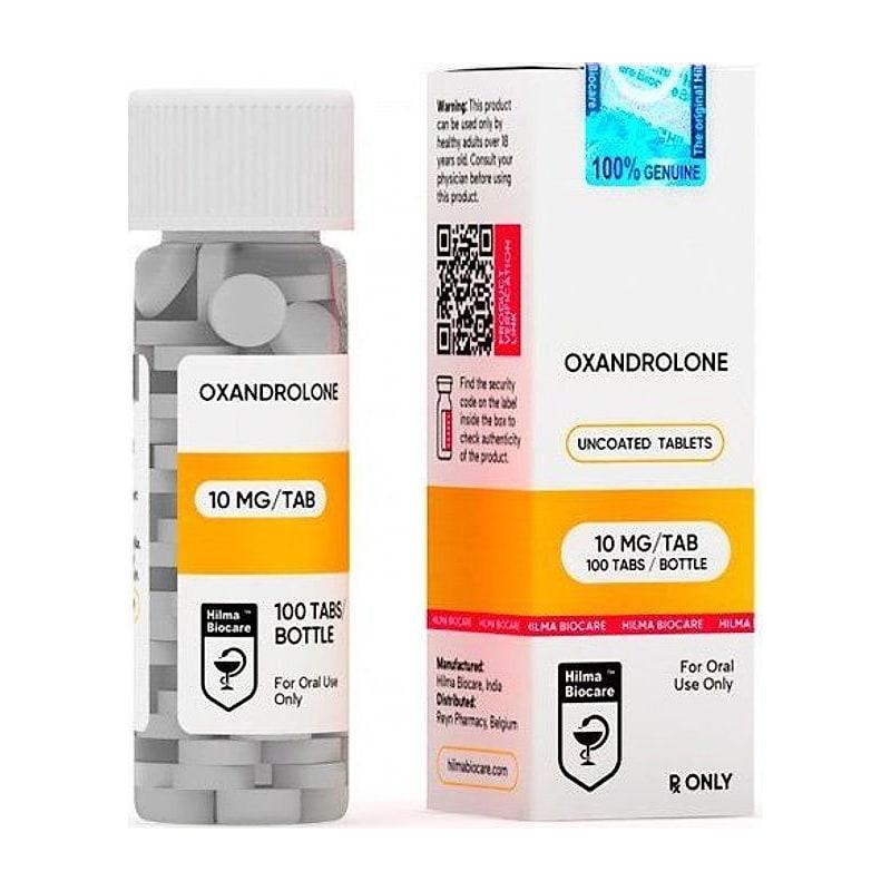
- Oxandrolone (C17α-methyl)
- Methandrostenolone/dianabol (C17α-methyl)
- Methyltestosterone (C17α-methyl; highest hepatotoxicity)
- Oxymetholone (C17α-methyl; highest hepatotoxicity in class)
Hepatotoxicity Mechanisms#
Three primary mechanisms operate in parallel:
1. BSEP inhibition → cholestatic injury (dominant mechanism):
- BSEP (bile salt export pump, ABCB11): hepatocyte canalicular membrane transporter; exports bile salts from hepatocyte into bile canaliculi
- C17α-alkylated steroids → competitive and non-competitive BSEP inhibition → intrahepatic bile salt accumulation → cholestasis
- Bile salts (particularly CDCA, LCA) → detergent effect on mitochondrial membrane → mPTP opening → necrosis
- Clinical phenotype: cholestatic hepatitis (elevated ALP, direct bilirubin, GGT with modest ALT rise)
2. CYP450 reactive metabolite formation → oxidative/immunologic injury:
- CYP3A4 metabolism of C17α-alkylated steroids → quinone, epoxide, and other reactive metabolites
- Reactive metabolites → form protein adducts → hepatocyte damage and potential immune-mediated DILI
- GSH depletion → oxidative stress → lipid peroxidation → hepatocellular necrosis
- Clinical phenotype: hepatocellular injury (marked ALT/AST elevation)
3. Direct mitochondrial toxicity:
- High intrahepatic steroid concentrations → electron transport chain disruption
- Decreased mitochondrial β-oxidation → lipid accumulation → steatosis/steatohepatitis
- Microvesicular steatosis (mitochondrial damage pattern) vs. macrovesicular (lipid flux disruption)
Biomarkers: Liver Enzyme Panels#
Standard liver enzyme monitoring in research:
| Biomarker | Type | What it reflects | Normal range (rats) |
|---|---|---|---|
| ALT (alanine aminotransferase) | Hepatocellular injury | Liver cell cytoplasmic damage | 17–70 U/L |
| AST (aspartate aminotransferase) | Hepatocellular + muscle | Less specific (also cardiac, skeletal) | 45–150 U/L |
| ALP (alkaline phosphatase) | Cholestatic marker | Bile canalicular damage | 60–200 U/L |
| GGT (gamma-glutamyl transferase) | Cholestatic marker | Bile duct and hepatocyte damage | 0–6 U/L |
| Direct bilirubin | Cholestatic marker | Impaired bilirubin excretion | <0.1 mg/dL |
| Total protein / albumin | Synthetic function | Liver synthetic capacity | Albumin 3.5–5.5 g/dL |
| PT/INR | Synthetic function | Clotting factor synthesis | PT ~9–13 sec |
Pattern interpretation:
- Hepatocellular pattern: ALT/AST elevated >5× ULN; ALP/GGT modestly elevated → CYP450 reactive metabolite or direct cell injury
- Cholestatic pattern: ALP/GGT/bilirubin elevated; ALT modestly elevated → BSEP inhibition dominant
- Mixed pattern: Both hepatocellular and cholestatic markers elevated → most common with C17α-alkylated steroids
R ratio (hepatocellular:cholestatic): R = (ALT/ULN) / (ALP/ULN)
- R >5: hepatocellular; R 2–5: mixed; R <2: cholestatic
Histopathology Spectrum#
C17α-alkylated steroids produce a distinctive histological spectrum:
1. Cholestasis:
- Canalicular bile plugs (bile thrombi in dilated bile canaliculi)
- Pericentral zone 3 predominance (highest CYP450 activity and most vulnerable to hepatocellular necrosis)
- Staining: H&E shows green-brown bile casts in canaliculi; confirmed by Hall's bilirubin stain
2. Peliosis hepatis:
- Blood-filled cystic spaces within hepatic parenchyma
- Pathognomonic for anabolic steroid exposure (especially C17α-alkylated)
- Mechanism: weakening of sinusoidal wall → rupture and blood-filled lakes
- May be silent radiologically (small lesions); severe cases → hepatic rupture risk
3. Hepatocellular necrosis:
- Pericentral (zone 3) coagulative necrosis in severe cases
- Lobular disarray, apoptotic bodies (councilman bodies)
4. Steatosis (lipid accumulation):
- Microvesicular: small lipid droplets within cytoplasm; nucleus central; indicates mitochondrial fatty acid oxidation impairment → more severe
- Macrovesicular: single large vacuole displacing nucleus; indicates lipid flux disruption; less severe
5. Hepatocellular carcinoma (HCC) — long-term:
- C17α-alkylated steroids associated with HCC in case reports (primarily oxymetholone, methyltestosterone)
- Mechanism: AR-mediated hepatocyte proliferation + genotoxic reactive metabolites
- Not characterized in typical 8–12 week research protocols; relevant for chronic exposure studies
Compound-Specific Hepatotoxicity Profiles#
| Compound | Relative hepatotoxicity | Dominant pattern | Key histology |
|---|---|---|---|
| Oxymetholone | Highest | Cholestatic + peliosis | Peliosis hepatis, canalicular cholestasis |
| Methyltestosterone | Very high | Cholestatic + hepatocellular | Cholestasis, mild necrosis |
| Methandrostenolone | High | Cholestatic | Canalicular cholestasis |
| Stanozolol | Moderate-high | Cholestatic + peliosis | Peliosis, bile plugs |
| Oxandrolone | Moderate | Hepatocellular + cholestatic | Steatosis, mild cholestasis; lowest in class |
Oxandrolone is generally considered the least hepatotoxic C17α-alkylated compound due to its 2-oxa ring modification (ring A oxygen substitution), which alters CYP3A4 metabolism and reduces reactive metabolite formation.
Hepatoprotective Strategies in Research#
1. TUDCA (tauroursodeoxycholic acid):
- Mechanism: replaces toxic bile salts in pool + anti-apoptotic + ER stress reduction
- Evidence: reduces ALT/AST and liver histology damage in C17α-steroid + TUDCA vs. steroid-alone rodent models
- Rodent dose: 250–500 mg/kg/day oral in drinking water
2. NAC (N-acetylcysteine):
- Mechanism: glutathione (GSH) precursor → replenishes hepatic GSH → quenches reactive metabolites
- Evidence: well-established for acetaminophen DILI (same GSH depletion mechanism); some data for anabolic steroid DILI
- Rodent dose: 150 mg/kg/day IP or 1–2% in drinking water
3. Silymarin (Milk Thistle):
- Mechanism: flavonolignan → membrane stabilization, antioxidant, anti-fibrotic
- Modest evidence for DILI mitigation; often used as positive control in hepatoprotection studies
4. Combined protocol (TUDCA + NAC):
- Rationale: TUDCA targets BSEP/cholestatic mechanism; NAC targets reactive metabolite/oxidative mechanism → complementary
- Research question: is the combination superior to either alone?
Research Protocol Design#
Hepatotoxicity dose-response study (rats)#
| Parameter | Specification |
|---|---|
| Model | Adult male Sprague-Dawley rats (250–300g) |
| Groups | 1. Vehicle, 2. Stanozolol 2 mg/kg/day, 3. Stanozolol 5 mg/kg/day, 4. Oxymetholone 5 mg/kg/day, 5. Oxandrolone 5 mg/kg/day |
| Route | Oral gavage × 8 weeks |
| Blood sampling | Baseline, week 2, week 4, week 6, week 8 |
| Endpoints | Serum ALT, AST, ALP, GGT, direct bilirubin, albumin; liver weight/body weight ratio; liver histology (H&E, Hall's bilirubin, Oil Red O); hepatic GSH; mitochondrial function (Complex I-III activity) |
Hepatoprotective co-treatment comparison#
| Parameter | Specification |
|---|---|
| Groups | 1. Vehicle, 2. Stanozolol 5 mg/kg/day, 3. Stanozolol + TUDCA 500 mg/kg/day, 4. Stanozolol + NAC 150 mg/kg/day, 5. Stanozolol + TUDCA + NAC |
| Duration | 6 weeks |
| Primary endpoint | ALT/AST area under curve (AUC) across 6 weeks |
| Secondary endpoints | Liver histology score (NAS/NAFLD score adaptation), BSEP expression by IHC, caspase-3 activation |
Key References#
-
Medina-Caliz I, et al. (2016). "Characterization and risk factors for anabolic steroid-induced drug-induced liver injury." Hepatology, 63(5):1473–1487.
-
Beuers U, et al. (1991). "Tauroursodeoxycholic acid stimulates hepatocellular exocytosis and vesicular secretion of bile acids in rats." Journal of Clinical Investigation, 87(4):1553–1561.
-
Neuman MG, et al. (1997). "Hepatic function in long-term androgenic-anabolic steroid users." Gut, 40(6):786–790.
Research Use Only: All compounds described are sold exclusively for in-vitro research and laboratory use. Not for unsupervised use. JuicedPharma does not provide medical advice.
Published by JuicedPharma Research Team · September 2026 · Category: Research Protocols & Science