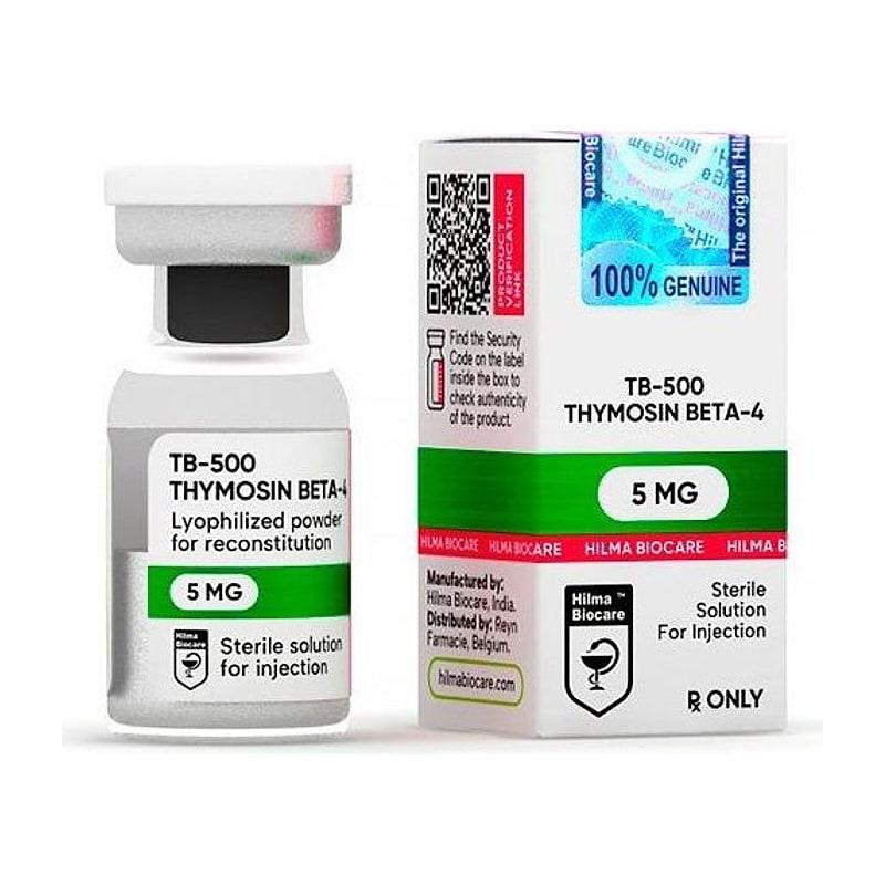
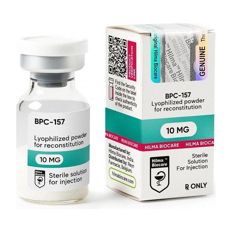
Research Disclaimer: TB-500 is sold exclusively for in-vitro and laboratory research use. Mechanistic information in this article is based on peer-reviewed preclinical and clinical research. This content does not constitute medical advice.
Understanding how a research compound works is the foundation of good experimental design. Researchers who know TB-500's mechanisms can predict which tissue models it will be most effective in, which outcome measures to track, and which confounds to control for. Those who treat it as a black box produce results that are harder to interpret and replicate.
This article is a mechanism-first deep dive into TB-500 (Thymosin Beta-4) — the actin biology, the signaling cascades, the cellular targets, and what each pathway means for tissue repair research.
Table of Contents#
- The Actin Cytoskeleton: Why It Matters
- G-Actin Sequestration: TB-500's Core Mechanism
- The G/F-Actin Ratio and Cell Migration
- Integrin-Linked Kinase (ILK) Activation
- IL-10 Upregulation: Anti-Inflammatory Action
- VEGF and Angiogenesis
- The LKKTETQ Binding Motif
- Mechanistic Differences from BPC-157
- Tissue-Specific Mechanistic Effects
- Research Design Implications
- Key Research References
The Actin Cytoskeleton: Why It Matters#
Before explaining how TB-500 works, it is worth establishing what the actin cytoskeleton is and why it is so central to tissue repair.
Every eukaryotic cell contains a dynamic network of protein filaments — the cytoskeleton — that determines the cell's shape, enables its movement, drives cell division, and transmits mechanical signals between the cell and its environment. Actin filaments are one of the three major cytoskeletal components (alongside microtubules and intermediate filaments).
Actin filaments are polymers of actin monomers. The dynamics of assembly and disassembly of these polymers — controlled by dozens of regulatory proteins — determines:
- Whether a cell can migrate: migrating cells extend protrusions (lamellipodia and filopodia) at their leading edge by rapidly polymerizing actin, then retract their trailing edge
- How fast a cell migrates: the rate of protrusion is directly tied to actin polymerization speed
- Whether a cell can squeeze through dense tissue: deformable cells with more globular actin traverse narrow spaces between other cells and matrix fibers more easily
In tissue repair, the speed and efficiency of cellular infiltration into the wound zone — fibroblasts laying down collagen, endothelial cells forming new vessels, keratinocytes re-covering the wound surface — is largely limited by cell migration capacity. TB-500 acts directly on this limiting factor.
G-Actin Sequestration: TB-500's Core Mechanism#
Actin exists in two interconvertible states:
- G-actin (globular actin) — the monomeric, soluble form. This is the building block.
- F-actin (filamentous actin) — the polymerized form. G-actin monomers associate end-to-end into long, double-helical filaments.
The conversion between G-actin and F-actin is regulated by a set of proteins including profilin, cofilin, capping proteins, Arp2/3, and the β-thymosins — the family to which Thymosin Beta-4 belongs.
TB-500 (Tβ4) sequesters G-actin. It binds directly to G-actin monomers in a 1:1 complex, holding them in the globular state and preventing them from adding to the growing end (barbed end) of F-actin filaments.
The sequestration-to-mobilization switch#
At first pass, sequestering G-actin sounds like it would reduce actin polymerization and slow cell migration. The opposite is often observed. Here is why:
In a resting cell with normal F-actin content, adding TB-500 sequesters more G-actin → reduces F-actin content slightly → creates a more deformable cell. When migration is triggered by a chemotactic signal, the cell releases G-actin from the Tβ4 complex → a burst of actin polymerization occurs at the leading edge → rapid protrusion formation.
TB-500 acts as a G-actin reservoir — buffering free G-actin during steady state and releasing it rapidly when the cell needs to move. This is why TB-500 promotes migration in activated (injury-sensing) cells while the effect on resting cells is subtler.
The G/F-Actin Ratio and Cell Migration#
The ratio of G-actin to F-actin is a quantifiable parameter in cell biology experiments. Higher G/F ratios (more globular, less filamentous actin) correlate with:
- Higher cell migration speed in scratch-assay models
- More lamellipodia formation at the leading edge
- Greater ability to pass through narrow pores in transwell assays (a model of migration through tissue)
- Less cell stiffness by atomic force microscopy
Studies measuring the G/F-actin ratio after TB-500 treatment find a consistent shift toward higher G/F — more free G-actin relative to polymerized F-actin — which matches the observed increase in migration.
Quantitative example from the literature#
Philp et al. (2004) demonstrated that endothelial cells treated with Tβ4 showed significantly increased migration distance in scratch assays, correlated with increased G-actin content. The mechanism was confirmed by competition: adding excess F-actin (which competes for Tβ4 binding) abolished the migration effect.
Integrin-Linked Kinase (ILK) Activation#
Beyond direct actin sequestration, TB-500 activates Integrin-Linked Kinase (ILK) — a scaffold and signaling protein at the integrin-cytoskeleton interface.
What is ILK?#
ILK sits at focal adhesion complexes — the points where integrins (cell surface receptors that bind the extracellular matrix) connect to the intracellular actin cytoskeleton. ILK is not primarily a kinase in the classical sense; its kinase domain is pseudokinase in many models. Rather, it functions as a scaffold that:
- Recruits and activates Akt (PKB) — promoting cell survival
- Activates GSK-3β phosphorylation — which promotes cell cycle progression
- Modulates integrin-mediated adhesion signaling — affecting how cells adhere to and migrate through the matrix
How TB-500 activates ILK#
Bock-Marquette et al. (2004, Nature) demonstrated that Tβ4 treatment of cardiac cells activates ILK. The mechanism involves Tβ4 binding to the ILK complex indirectly through PI3-kinase activation — the G-actin sequestration and cytoskeletal effects are upstream, and ILK activation appears to require the cytoskeletal changes that Tβ4 induces.
Functional consequence: cell survival#
ILK activation is a survival signal. In ischemic cardiac tissue, where cardiomyocytes are dying from ATP depletion, Tβ4-mediated ILK activation promotes Akt phosphorylation, which:
- Inhibits pro-apoptotic proteins (BAD, caspase-9)
- Maintains mitochondrial membrane potential
- Reduces cardiomyocyte death in the infarct border zone
This is the mechanistic basis for TB-500's most clinically relevant effect — cardiac protection.
IL-10 Upregulation: Anti-Inflammatory Action#
A third mechanistic pathway of TB-500 is the upregulation of Interleukin-10 (IL-10), a key anti-inflammatory cytokine.
IL-10's role in tissue repair#
Inflammation is necessary for tissue repair — it clears debris, recruits immune cells, and initiates the proliferative response. However, excessive or prolonged inflammation impairs repair by:
- Damaging healthy tissue adjacent to the injury zone
- Promoting fibrosis (scar formation) over regeneration
- Inhibiting stem cell activity in the repair zone
IL-10 is an anti-inflammatory cytokine that counter-regulates pro-inflammatory signals (TNF-α, IL-1β, IL-6). It is associated with the transition from acute inflammation to the proliferative (repair) phase of wound healing.
TB-500 and IL-10#
Studies in wound healing and cardiac ischemia models show that TB-500 treatment increases local IL-10 levels. The mechanism for this is not fully characterized — it may be indirect (through the macrophage polarization shift that TB-500 appears to promote) or direct (through transcriptional effects on cytokine genes).
The practical consequence: in models where excessive inflammation is a barrier to repair — chronic wounds, post-infarction remodeling, inflammatory bowel conditions — TB-500's IL-10 upregulation may be as important as its direct migration effects.
VEGF and Angiogenesis#
TB-500 promotes Vascular Endothelial Growth Factor (VEGF) expression in several model systems, driving angiogenesis — the formation of new blood vessels.
Adequate blood supply to the healing tissue is essential for:
- Oxygen and nutrient delivery to proliferating cells
- Removal of metabolic waste from the repair zone
- Immune cell trafficking into and out of the wound
TB-500 promotes endothelial cell migration (via G-actin sequestration) and proliferation (via ILK/Akt) while also upregulating VEGF, which acts in an autocrine/paracrine loop to further stimulate endothelial recruitment. The combined effect is accelerated vascularization of the healing zone.
In the cardiac research context, this angiogenic effect is particularly relevant: post-infarction tissue is chronically ischemic partly due to coronary microvessel loss. Restoring microvasculature with VEGF-driven angiogenesis is a key therapeutic target, and TB-500's angiogenic activity is one reason the compound was developed for cardiac indications.
The LKKTETQ Binding Motif#
The Thymosin Beta-4 sequence responsible for G-actin binding has been localized to a specific segment: LKKTETQ (amino acids 17–23). This tetrapeptide/heptapeptide motif is the minimal actin-binding unit of Tβ4.
Research using synthetic LKKTETQ peptide alone has confirmed that this motif retains:
- G-actin sequestration activity
- Promotion of endothelial cell migration
- Some wound healing acceleration
However, the isolated LKKTETQ motif lacks the full biological activity of complete Tβ4. Specifically:
- ILK activation has not been consistently reproduced with LKKTETQ alone
- Anti-inflammatory effects (IL-10) are weaker or absent
- Cardiac protection effects require the full-length sequence
This is why research-grade TB-500 should be the full 43-amino-acid sequence — not just the LKKTETQ fragment — for experiments targeting the full mechanistic profile.
Mechanistic Differences from BPC-157#
| Mechanism | TB-500 | BPC-157 |
|---|---|---|
| Actin dynamics | G-actin sequestration (direct) | No direct actin effect |
| Cell migration | Via cytoskeletal remodeling | Via FAK-paxillin signaling |
| Survival signaling | ILK → Akt | GHR upregulation |
| Anti-inflammatory | IL-10 upregulation | Eicosanoid modulation + NO |
| Angiogenesis | VEGF upregulation | VEGF upregulation |
| GI protection | No specific mechanism | Strong (gastric origin) |
| CNS effects | Limited data | Dopaminergic normalization |
The two pathways for cell migration (G-actin sequestration vs. FAK-paxillin signaling) are upstream-different — activating different nodes of the migration machinery. This is the mechanistic rationale for their combination in stacking protocols.
Tissue-Specific Mechanistic Effects#
Cardiac tissue#
ILK activation → Akt → anti-apoptotic signaling in cardiomyocytes. VEGF-driven coronary angiogenesis. Epicardial progenitor mobilization (via Smart et al. 2007 pathway). IL-10 reduction of post-infarction inflammatory remodeling.
Skin / wound#
G-actin sequestration drives keratinocyte migration for re-epithelialization. VEGF supports dermal neovascularization. IL-10 prevents excessive fibrosis, allowing regenerative rather than scar-forming healing.
Muscle#
Satellite cell activation (the muscle-specific stem cells) appears to be promoted by TB-500, though this is less well-characterized than the cardiac and skin evidence. Reduced fibrosis in muscle crush models suggests the IL-10 pathway is active in this context.
Connective tissue / tendon#
Fibroblast migration is driven by G-actin sequestration. Collagen deposition occurs downstream of fibroblast infiltration. Less evidence here than for BPC-157's tendon-specific studies — TB-500 in connective tissue is a secondary indication relative to cardiac and skin.
Research Design Implications#
Understanding TB-500's mechanisms directly informs experimental design:
Endpoint selection:
- For G-actin sequestration studies: measure G/F-actin ratios by DNase I inhibition assay or differential centrifugation
- For ILK/Akt pathway: Western blot for pAkt (Ser473) and pGSK-3β (Ser9) in treated cells
- For IL-10: ELISA on conditioned medium or tissue homogenate
- For migration: scratch assay or transwell assay; image at 12–24h intervals
- For angiogenesis: endothelial tube formation assay; VEGF ELISA
Timing:
- Migration effects are rapid (hours to days) — capture early time points
- Angiogenic effects are slower (days to weeks) — allow adequate study duration
- Anti-apoptotic effects peak in the acute post-injury window (6–24h) — dose early relative to injury
Dose range:
- G-actin sequestration is a biochemical (stoichiometric) interaction — higher doses up to saturation of the free G-actin pool are meaningful
- Research doses of 2–5 mg/kg total (not per kg daily) in preclinical models reflect this higher mass requirement compared to BPC-157
Key Research References#
-
Bock-Marquette I, et al. (2004). "Thymosin beta4 activates integrin-linked kinase and promotes cardiac cell migration, survival and cardiac repair." Nature, 432(7016):466–472.
-
Smart N, et al. (2007). "Thymosin beta4 induces adult epicardial progenitor mobilization and neovascularization." Nature, 445(7124):177–182.
-
Philp D, et al. (2004). "Thymosin beta 4 and a synthetic tetrapeptide AcSDKP promote endothelial cell adhesion and migration." FASEB Journal, 18(6):760–762.
-
Sosne G, et al. (2007). "Thymosin beta4 promotes corneal wound healing and modulates inflammatory mediators in vivo." Experimental Eye Research, 84(1):166–172.
-
Huff T, et al. (2004). "Beta-thymosins, small acidic peptides with multiple functions." International Journal of Biochemistry & Cell Biology, 36(11):2023–2035.
-
Goldstein AL, et al. (2005). "Thymosin β4: a novel molecular target for antitumor strategies." Nature Reviews Cancer, 5(1):67–76.
-
Crockford D, et al. (2010). "Effect of thymosin beta-4 on human keratinocyte migration." Wound Repair and Regeneration, 18(1):48–54.
Research Use Only: TB-500 and all products referenced are sold exclusively for in-vitro research and laboratory use. Not for human consumption. JuicedPharma does not provide medical advice.
Published by JuicedPharma Research Team · September 2026 · Category: Peptides & Recovery