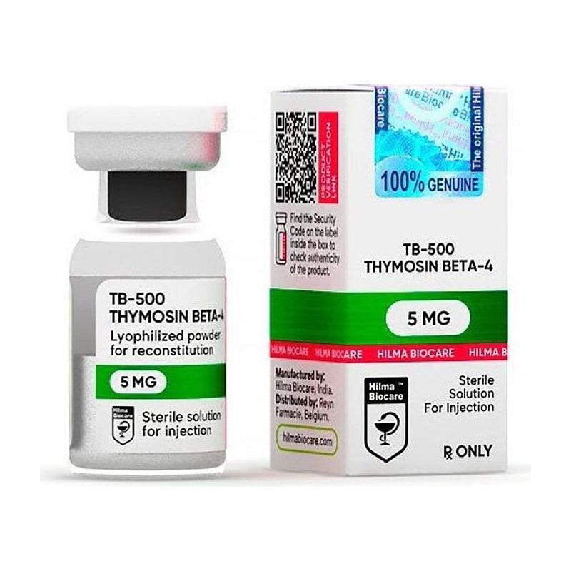
Research Disclaimer: TB-500 is sold exclusively for in-vitro and laboratory research use. This article covers the molecular biology underlying TB-500 research. It does not constitute medical advice.
Actin is one of the most abundant proteins in eukaryotic cells and one of the most studied in cell biology. Yet the role of actin-sequestering proteins — particularly the β-thymosin family to which TB-500 belongs — is not always well understood by researchers approaching TB-500 from a clinical or pharmacological angle.
This article bridges that gap. Understanding actin dynamics is not optional for TB-500 research — it is the mechanism. Researchers who understand the G/F-actin equilibrium, the profilin/thymosin competition, and the relationship between cytoskeletal state and cell behavior will design better experiments and interpret their results more accurately.
Table of Contents#
- Actin: Structure and Function
- The G-Actin/F-Actin Equilibrium
- Actin Treadmilling and Cell Motility
- The β-Thymosin Family: Where TB-500 Belongs
- How TB-500 Sequesters G-Actin: Binding Mechanics
- The Profilin-Thymosin Competition
- Downstream Consequences of G-Actin Sequestration
- Measuring G/F-Actin Ratios in Research
- In Vitro Assay Designs for TB-500 Actin Research
- Key Research References
Actin: Structure and Function#
Actin is a 42 kDa globular protein that serves as the monomer building block of actin filaments. It was first purified from skeletal muscle in the 1940s (Straub, 1942) and has since been identified in every eukaryotic cell type studied.
The mature actin protein:
- Is encoded by six isoforms in mammals (α-cardiac, α-skeletal, α-smooth, β-cytoplasmic, γ-cytoplasmic, γ-smooth)
- Binds one molecule of ATP (or ADP after hydrolysis) and one Ca²⁺ or Mg²⁺ ion per monomer
- Has a molecular weight of 41.7 kDa in its monomeric form
- Contains a hydrophobic cleft that mediates both monomer-monomer polymerization contacts and binding by regulatory proteins
The β and γ cytoplasmic isoforms are the predominant forms in non-muscle cells — the forms most relevant to TB-500 research in migration, wound healing, and repair models.
The G-Actin/F-Actin Equilibrium#
In cells, actin exists in a dynamic equilibrium between two states:
G-actin (globular actin)#
- Monomeric, soluble form
- ATP-bound (G-actin-ATP is the predominant free form)
- Concentration in most cells: 50–200 µM
- Functions: pool of building blocks for polymerization; binds regulatory proteins
F-actin (filamentous actin)#
- Polymerized form — a right-handed double helix of G-actin monomers
- ADP-bound (ATP is hydrolyzed during/after polymerization)
- Provides structural rigidity, force generation, and tracks for myosin motors
- Half-life of individual filaments: seconds to minutes (highly dynamic)
The ratio of G-actin to F-actin (G/F ratio) is not fixed — it varies by:
- Cell type (neurons have different ratios than fibroblasts)
- Cell state (migrating vs. resting vs. dividing cells have very different ratios)
- Regulatory protein concentrations (thymosins, profilins, cofilin, Arp2/3)
- Extracellular signals (growth factors, chemokines shift the ratio within minutes)
TB-500 shifts this ratio toward G-actin — more free monomers, fewer polymerized filaments per unit volume.
Actin Treadmilling and Cell Motility#
Cell migration depends on actin treadmilling — the asymmetric addition and removal of G-actin from opposite ends of F-actin filaments:
- Barbed end (plus end): the "fast" end where G-actin-ATP monomers are added. Located at the leading edge of migrating cells.
- Pointed end (minus end): the "slow" end where ADP-actin is released back into the G-actin pool. Located toward the cell body.
In a migrating cell:
- Arp2/3 complex nucleates new filaments at the barbed end → branched F-actin network forms → the cell edge protrudes outward (lamellipodia)
- Cofilin severs and depolymerizes filaments at the pointed end → ADP-actin released → ATP exchanged → G-actin-ATP pool replenished
- Net result: actin "flows" from the pointed end to the barbed end, pushing the cell membrane forward
For this cycle to run efficiently, the G-actin-ATP pool must be adequate. If the pool is depleted (too much F-actin, not enough G-actin available), treadmilling slows and migration halts.
TB-500 maintains the G-actin-ATP pool by sequestering monomers and keeping them available (and properly nucleotide-exchanged) for rapid deployment to barbed ends when migration signals arrive.
The β-Thymosin Family: Where TB-500 Belongs#
The β-thymosin proteins are a family of small, intrinsically disordered peptides with the shared function of G-actin sequestration. The family includes:
| Protein | Length | Expression |
|---|---|---|
| Thymosin β4 (TB-500) | 43 aa | Ubiquitous (highest in platelets) |
| Thymosin β10 | 43 aa | Embryonic development, some adult tissues |
| Thymosin β15 | 44 aa | Prostate, some cancer lines |
Thymosin β4 (TB-500) is the most abundant and widely expressed family member in adult mammalian tissue. Intracellular concentrations of Tβ4 have been measured at 300–550 µM in platelets — high enough to sequester a substantial fraction of the total G-actin pool.
The conservation of this protein family across evolution (homologs exist in sea urchins, Drosophila, and C. elegans) underscores the fundamental importance of G-actin sequestration in basic cell biology.
How TB-500 Sequesters G-Actin: Binding Mechanics#
The Tβ4-G-actin interaction is well-characterized structurally:
Binding affinity#
The dissociation constant (Kd) for the Tβ4-G-actin complex has been measured at approximately 0.5–2 µM. Given that free G-actin concentrations in cells are typically 10–100 µM (above the critical concentration for polymerization), and intracellular Tβ4 concentrations are in the range of 100–500 µM (in platelets), the math indicates that a large fraction of G-actin in Tβ4-rich cells is bound.
Binding site#
Tβ4 binds G-actin through two contact regions:
- The N-terminal region (residues 1–16): minor contact, includes the Ac-Ser at position 1 (N-acetylation is important for optimal binding)
- The central helix (residues 17–28, the LKKTETQ motif and surrounding helix): the primary binding region. This helix inserts into the hydrophobic cleft on the actin surface.
The binding is not covalent — it is a reversible non-covalent complex maintained by hydrophobic and electrostatic contacts.
Binding prevents polymerization#
The Tβ4 binding site on actin overlaps with the barbed-end contact surface used for filament elongation. By occupying this site, Tβ4 physically prevents G-actin from adding to the growing barbed end — hence "sequestration."
The Profilin-Thymosin Competition#
Thymosin β4 is not the only G-actin-binding protein in cells. Profilin is a major competitor — and the profilin-thymosin balance determines whether G-actin is sequestered (thymosin wins) or channeled toward filament elongation (profilin wins).
How profilin works#
Profilin binds G-actin at the barbed-end surface (same site as thymosins) but, unlike thymosins, profilin-actin complexes CAN be added to barbed ends. Profilin acts as a "delivery vehicle" — it accompanies G-actin to the growing barbed end, adds the actin monomer, and dissociates. Profilin also catalyzes nucleotide exchange (ADP → ATP) on released actin monomers, maintaining the active ATP-bound pool.
The tug-of-war#
Profilin has a higher affinity for G-actin than thymosin (Kd ≈ 0.1 µM for profilin vs. ≈1 µM for Tβ4). However, thymosins are present at much higher concentrations than profilin (Tβ4 can be 10–50× more concentrated than profilin in some cell types).
The cell uses the concentration advantage of thymosins and the affinity advantage of profilin to set the G-actin buffering point — determining how much G-actin is available for fast polymerization vs. how much is held in reserve.
When TB-500 is added exogenously, it shifts this equilibrium toward higher thymosin/actin ratios → more buffered G-actin → paradoxically more efficient barbed-end elongation when migration signals arrive (because a large burst release of thymosin-bound G-actin can be triggered by local signaling).
Downstream Consequences of G-Actin Sequestration#
The actin cytoskeleton is not only a structural component — it is a signaling hub. Changes in G/F-actin ratio directly regulate gene transcription:
MRTF-A/SRF transcriptional pathway#
The transcriptional coactivator MRTF-A (Myocardin-Related Transcription Factor A) is sequestered in the cytoplasm when bound to G-actin. When G-actin levels decrease (more F-actin, less free G-actin), MRTF-A is released → translocates to the nucleus → activates SRF (Serum Response Factor) → drives transcription of:
- Actin isoforms (cytoskeletal actin genes)
- Smooth muscle actin (αSMA)
- Connective tissue growth factor (CTGF)
- Integrins
TB-500 increases free G-actin → sequesters MRTF-A → reduces SRF activity. This has counterintuitive consequences for collagen production and fibrosis — TB-500 may partially suppress excessive fibrosis by keeping MRTF-A cytoplasmic.
This pathway provides a mechanistic link between TB-500's actin sequestration and the observed anti-fibrotic effects in muscle repair models.
Measuring G/F-Actin Ratios in Research#
TB-500 studies that target the actin mechanism should include direct G/F-actin ratio measurements:
DNase I inhibition assay#
G-actin inhibits DNase I; F-actin does not. After cell lysis under conditions that preserve the G/F ratio, DNase I activity correlates inversely with G-actin content. This assay is sensitive but requires careful lysis conditions.
Differential centrifugation#
Cell lysates spun at high speed (100,000 × g) pellet F-actin; G-actin remains in the supernatant. Pellet and supernatant fractions are run on SDS-PAGE and quantified by densitometry. Simple and reproducible; requires ultracentrifuge access.
Phalloidin / G-actin antibody immunofluorescence#
Fluorescent phalloidin (binds F-actin only) and anti-G-actin antibody staining can be visualized and quantified by confocal microscopy in fixed cells. Allows spatial information about where in the cell the ratio changes occur.
In Vitro Assay Designs for TB-500 Actin Research#
Scratch (wound) assay#
- Culture cells to confluence on 6-well plates
- Create linear scratch with a pipette tip
- Add TB-500 at target concentration (typically 100–1000 ng/ml for in vitro)
- Image at 0, 6, 12, 24h
- Measure wound closure rate (area or leading edge migration distance)
Transwell migration assay#
- Cells seeded in upper chamber of transwell insert (8 µm pores)
- TB-500 added to lower chamber as chemoattractant, or both chambers
- Fix and stain cells on the underside of the membrane at 24–48h
- Count migrated cells per field
F-actin staining (phalloidin)#
- Fix cells (4% paraformaldehyde) after TB-500 treatment
- Stain with fluorescent phalloidin (Alexa Fluor 488-phalloidin is standard)
- Compare F-actin content and architecture (cortical vs. stress fibers) between TB-500 treated and control cells
- Quantify mean fluorescence intensity per cell
G/F-actin ratio quantification#
- Combine differential centrifugation with Western blot for actin
- Compare actin in supernatant (G) vs. pellet (F) fractions
- Calculate G/F ratio for treated vs. control cells at multiple time points post-TB-500 addition
Key Research References#
-
Huff T, et al. (2004). "Beta-thymosins, small acidic peptides with multiple functions." International Journal of Biochemistry & Cell Biology, 36(11):2023–2035.
-
Safer D, et al. (1991). "Thymosin beta 4 abolishes in the presence of profilin the barbed end capping activities of beta-thymosins." Journal of Cell Biology, 115(2):289–296.
-
Hertzog M, et al. (2004). "The beta-thymosin/WH2 domain; structural basis for the switch from inhibition to promotion of actin assembly." Cell, 117(5):611–623.
-
Pollard TD, Borisy GG. (2003). "Cellular motility driven by assembly and disassembly of actin filaments." Cell, 112(4):453–465.
-
Philp D, et al. (2004). "Thymosin beta 4 and a synthetic tetrapeptide AcSDKP promote endothelial cell adhesion and migration." FASEB Journal, 18(6):760–762.
-
Bock-Marquette I, et al. (2004). "Thymosin beta4 activates integrin-linked kinase." Nature, 432(7016):466–472.
-
Miralles F, et al. (2003). "Actin dynamics control SRF activity by regulation of its coactivator MAL." Cell, 113(3):329–342.
Research Use Only: TB-500 and all products referenced are sold exclusively for in-vitro research and laboratory use. Not for human consumption. JuicedPharma does not provide medical advice.
Published by JuicedPharma Research Team · September 2026 · Category: Research Protocols & Science